 GenMI-LabMBZUAI
PRIMED
When Iteration Cannot Help: Single-Pass Missing-Modality MRI Synthesis and What Actually Limits It
*Equal first authors
Synthesise any missing MRI contrast from the others.
One network evaluation per volume.
28.56 dB on BraTS-GLI in 0.15 s.
01 / ABSTRACT
Abstract
Missing-modality MRI synthesis predicts an unavailable contrast from the remaining acquisitions, for which diffusion-based approaches often require hundreds or thousands of network evaluations per volume. We investigate when such iterative refinement can help in informed-prior bridges and show that, when the source state is a deterministic function of the conditioning, subsequent states cannot provide additional target information. However, stochastic training states expose the network to information unavailable during inference, creating a mismatch between training and deployment. To address this, we introduce PRIMED, a single-pass displacement regression framework that trains exclusively at the source endpoint and synthesises the missing contrast with one network evaluation. A single shared network handles all four target contrasts using a fixed 3D Haar wavelet representation. On BraTS-GLI, PRIMED achieves 28.56 dB PSNR and 0.944 SSIM in 0.152 s per volume, exceeding its two-evaluation bridge by 0.596 dB at half the runtime and achieving higher PSNR than thousand-step cWDM at comparable SSIM, with a 1{,}053\times speedup. Its effectiveness is further validated on unseen BraTS cohorts and a separately trained IXI setting. Beyond synthesis performance, we investigate the information limits of missing-modality reconstruction, finding that recoverability varies nearly fourfold across target contrasts and anatomical regions. Code is available at https://github.com/genmilab/PRIMED.
02 / METHOD
Method
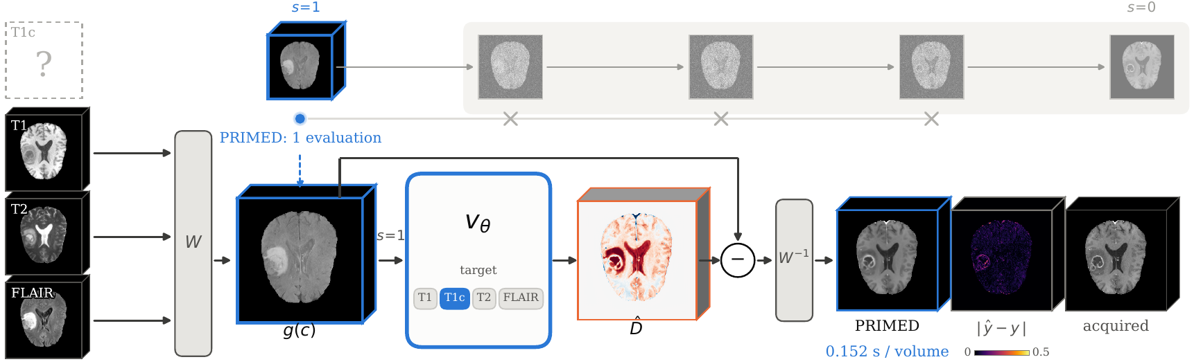
- Representation. A fixed, lossless one-level 3D Haar transform W maps each 224 × 224 × 160 volume to eight subbands of 112 × 112 × 80. The low-pass band is divided by 3.
- Informed prior. g(c) is the mean of the encoded acquired contrasts. It depends on the inputs alone, so the network can compute it exactly.
- Network. vθ is a five-level 3D U-Net (MONAI
DiffusionModelUNet, 97.7M parameters), convolutional except for one self-attention block in its bottleneck. Its 48 input channels are g(c) twice, a learned 8-channel embedding of the target contrast and the three encoded acquired contrasts. - Training. Mean squared error on the displacement D = g(c) − W y at the source state s = 1. No noise is injected.
- Inference. One network evaluation followed by the inverse transform:
$$\hat{y} = W^{-1}\big(g(c) - v_\theta(g(c), 1, c)\big), \qquad g(c) = \tfrac{1}{3}\textstyle\sum_{m} W c_m .$$
03 / RESULTS
Results
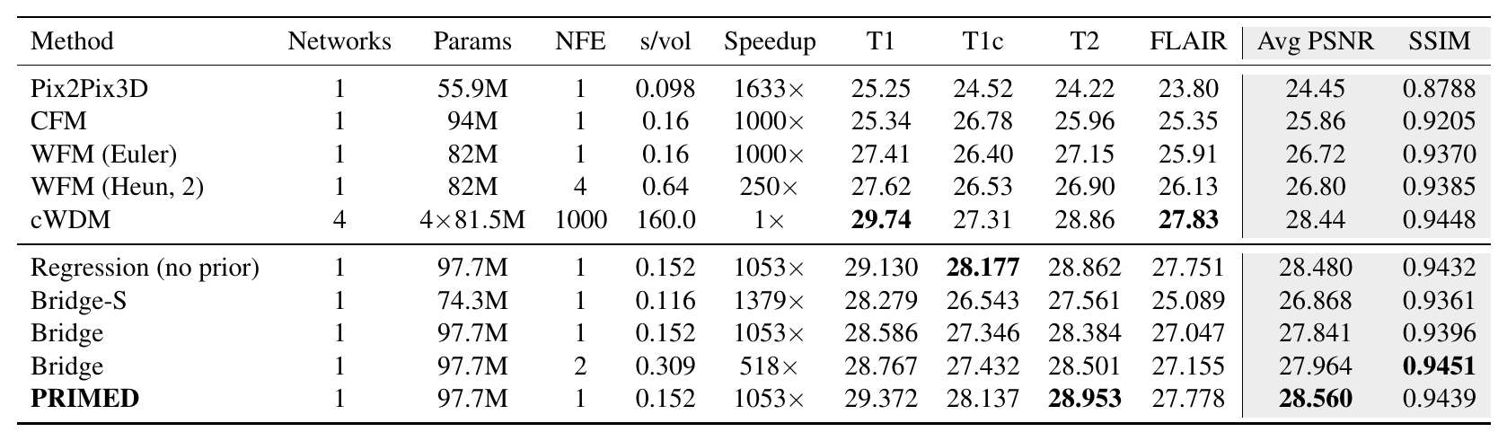
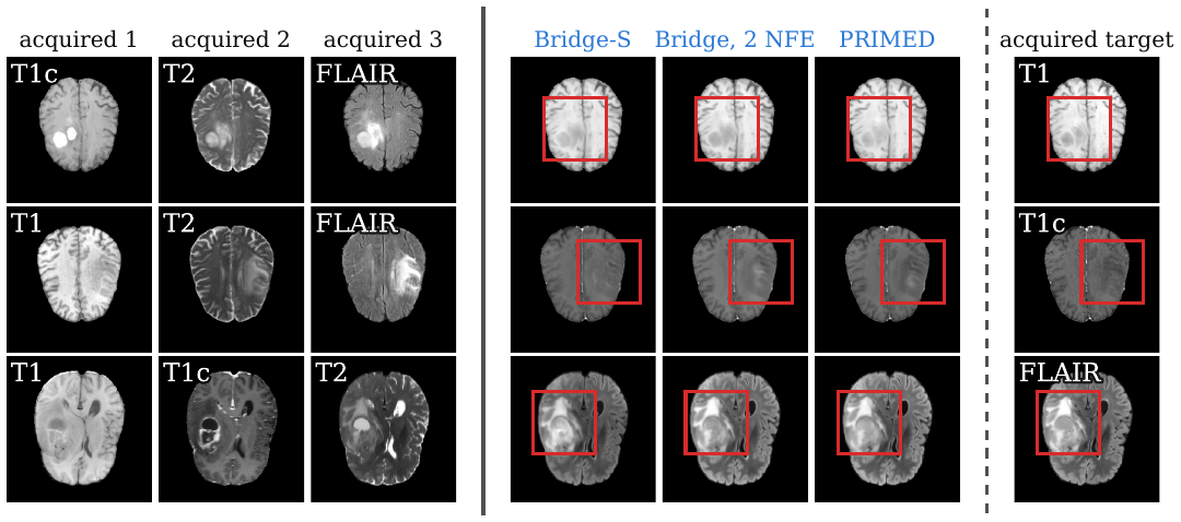
Whole-volume PSNR (dB) per target contrast and SSIM on the 216 held-out BraTS 2023 GLI subjects (864 cases), with one network for all four targets, as reported in the paper:
| Networks | NFE | Params | s / volume | T1 | T1c | T2 | FLAIR | Avg | SSIM |
|---|---|---|---|---|---|---|---|---|---|
| 1 | 1 | 97.7M | 0.152 | 29.372 | 28.137 | 28.953 | 27.778 | 28.560 | 0.9439 |
NFE is the number of network evaluations per volume. Timing is per volume on one NVIDIA RTX PRO 6000 Blackwell, including the inverse transform.
Inside the brain and the tumour regions
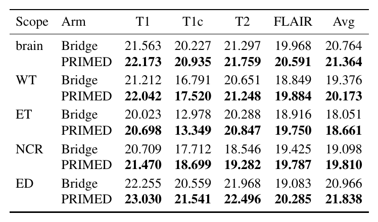
PSNR (dB), averaged over the four targets:
| Brain | Whole tumour | Enhancing tumour | Necrotic core | Oedema |
|---|---|---|---|---|
| 21.364 | 20.173 | 18.661 | 19.810 | 21.838 |
How much of each target the inputs determine varies by contrast and region. The recoverability statistic ρ is 0.632–0.740 over the whole brain, but 0.355, 0.188, 0.571 and 0.508 inside enhancing tumour for the T1, T1c, T2 and FLAIR targets.
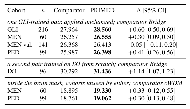
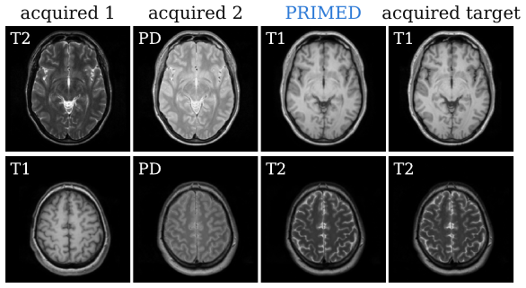
Unseen cohorts. The GLI-trained model applied unchanged, and a second model trained on IXI:
| Cohort | Subjects | PSNR (dB) |
|---|---|---|
| BraTS-MEN, training release | 60 | 26.555 |
| BraTS-MEN, validation release | 141 | 26.413 |
| BraTS-PED | 99 | 26.398 |
| IXI (trained on IXI) | 96 | 31.436 |
Fewer acquired contrasts. primed_brats_avail is fine-tuned for 8k steps with random input availability; empty input slots receive the prior. Mean PSNR (dB) over all four targets and every subset of the inputs:
| Acquired contrasts | primed_brats, zero-shot |
primed_brats_avail |
|---|---|---|
| three | 28.56 | 28.36 |
| two | 25.40 | 27.35 |
| one | 22.37 | 25.97 |
Misregistered or thick-slice inputs. primed_brats_robust is fine-tuned for 8k steps with these perturbations applied to the inputs. Mean PSNR (dB):
| Inputs | primed_brats |
primed_brats_robust |
|---|---|---|
| registered | 28.56 | 28.44 |
| one input misregistered by 1 mm, 1° | 27.67 | 27.89 |
| one input misregistered by 2 mm, 2° | 26.60 | 27.28 |
| one input misregistered by 4 mm, 4° | 25.10 | 26.47 |
| all inputs misregistered by 2 mm, 2° | 23.88 | 24.71 |
| inputs from 3 mm slices | 26.67 | 27.58 |
| inputs from 5 mm slices | 24.66 | 26.82 |
From the paper
04 / CODE
Code and usage
Installation
git clone https://github.com/genmilab/PRIMED.git
cd PRIMED
conda create -n primed python=3.10 -y && conda activate primed
pip install -e .
The code needs Python 3.9 or later, PyTorch 2.1 or later, MONAI 1.4 or later and einops. It was tested with MONAI 1.4.0.
Pretrained Weights
Download the checkpoints from Hugging Face:
pip install -U huggingface_hub
hf download adidukre/PRIMED --local-dir weights
cd weights && sha256sum -c SHA256SUMS
| File | Training data | Synthesises |
|---|---|---|
primed_brats.safetensors |
BraTS 2023 GLI, 1032 subjects | any of T1n, T1c, T2w, T2f from the other three |
primed_brats_avail.safetensors |
as primed_brats, plus random input availability |
any of the four from one, two or three acquired contrasts |
primed_brats_robust.safetensors |
as primed_brats, plus misregistration and thick-slice augmentation |
any of the four from three possibly misregistered or thick-slice contrasts |
primed_ixi.safetensors |
IXI, 477 subjects | any of T1, T2, PD from the other two |
Each file holds the network weights only. The architecture is read from the weights, so no config is needed for inference.
Data
BraTS. Download the BraTS 2023 adult glioma (GLI) training data from the challenge page and keep its folder layout:
/path/to/ASNR-MICCAI-BraTS2023-GLI-Challenge-TrainingData/
└── BraTS-GLI-00000-000/
    ├── BraTS-GLI-00000-000-t1n.nii.gz
    ├── BraTS-GLI-00000-000-t1c.nii.gz
    ├── BraTS-GLI-00000-000-t2w.nii.gz
    ├── BraTS-GLI-00000-000-t2f.nii.gz
    └── BraTS-GLI-00000-000-seg.nii.gz
Volumes must be co-registered, skull-stripped and on the 1 mm BraTS grid of 240 × 240 × 155 voxels, as BraTS releases them. Preprocessing runs on the fly: - clip to the 0.1 and 99.9 percentiles of the volume; - rescale to [0, 1]; - pad the depth from 155 to 160 slices; - crop the in-plane axes from 240 to 224.
IXI. Arrange the IXI dataset as /path/to/IXI/<subject>/<subject>-{T1,T2,PD}.nii.gz, with the three contrasts co-registered and resampled to the same 240 × 240 × 155, 1 mm grid. The IXI volumes are not skull-stripped.
splits/ holds the subject lists used in the paper:
splits/
├── brats_gli.json           # 1032 train / 219 val; 3 val subjects select checkpoints, the other 216 are the test set
├── brats_men_60.txt         # BraTS-MEN training release, first 60 subjects
├── brats_men_val_141.txt    # BraTS-MEN validation release
├── brats_ped_99.txt         # BraTS-PED training release
└── ixi.json                 # 477 train / 100 val; 4 val subjects select checkpoints, the other 96 are the test set
Inference
Synthesise a missing contrast from NIfTI files:
python synthesize.py \
  --weights weights/primed_brats.safetensors \
  --target t1c \
  --inputs t1n=/path/to/subject-t1n.nii.gz t2w=/path/to/subject-t2w.nii.gz t2f=/path/to/subject-t2f.nii.gz \
  --output /path/to/subject-t1c-synthesised.nii.gz
| Model | Contrast names (case-insensitive) |
|---|---|
| BraTS | t1n, t1c, t2w, t2f (aliases such as t1ce and flair also work) |
| IXI | t1, t2, pd |
The output is on the grid of the inputs, with intensities in the normalised [0, 1] range. With primed_brats_avail.safetensors, any one or two acquired contrasts may be given; the empty input slots are filled with their mean.
From Python
import torch
from primed import load_model
from primed.data import load_volume

device = "cuda" if torch.cuda.is_available() else "cpu"
model = load_model("weights/primed_brats.safetensors", device)
inputs = torch.stack(
    [load_volume(f"/path/to/subject-{m}.nii.gz") for m in ("t1n", "t2w", "t2f")]
).unsqueeze(0).to(device)
t1c = model.synthesize(inputs.mean(dim=1), inputs, torch.tensor([1], device=device))
The target class follows the contrast order T1n, T1c, T2w, T2f for BraTS and T1, T2, PD for IXI.
Inference needs about 5 GB of GPU memory. It also runs on the CPU (--device cpu), at about 25–30 s per volume with 16 threads.
Evaluation
Score the 216 held-out BraTS-GLI subjects and print the per-target table:
python evaluate.py \
  --config configs/brats_primed.yaml \
  --weights weights/primed_brats.safetensors \
  --output results/primed_brats_test.jsonl
Per-case results are written as JSON lines: whole-volume PSNR on the 224 × 224 × 155 crop, PSNR inside the brain and inside each tumour region, and 3D SSIM with a 7³ window.
| Option | Effect |
|---|---|
--num_inputs 2 / --num_inputs 1 |
every subset of fewer acquired contrasts (use primed_brats_avail.safetensors) |
--fill zero |
fill empty input slots with zeros instead of the mean of the acquired contrasts |
--split all --data_root DIR |
score another cohort |
--subjects FILE |
restrict scoring to the subjects listed in a file |
--limit N |
score only the first N subjects |
--device cpu |
run on the CPU |
For example, the 60 BraTS-MEN subjects:
python evaluate.py --config configs/brats_primed.yaml \
  --weights weights/primed_brats.safetensors \
  --split all --data_root /path/to/BraTS-MEN-Train --subjects splits/brats_men_60.txt \
  --output results/primed_brats_men60.jsonl
For IXI, use --config configs/ixi_primed.yaml with primed_ixi.safetensors.
Training
Set data_root, output_dir and, where used, init_weights in the config, then run from the repository root:
python train.py --config configs/brats_primed.yaml
torchrun --nproc_per_node=4 train.py --config configs/brats_primed.yaml
| Config | Initialisation | Augmentation | Released as |
|---|---|---|---|
configs/brats_primed.yaml |
init_weights (see below) |
random flips | primed_brats |
configs/brats_primed_avail.yaml |
primed_brats.safetensors |
flips, random input availability | primed_brats_avail |
configs/brats_primed_robust.yaml |
primed_brats.safetensors |
flips, misregistration and thick slices | primed_brats_robust |
configs/ixi_primed.yaml |
random | random flips | primed_ixi |
| Setting | Value |
|---|---|
| Optimiser | AdamW, weight decay 0, gradient clip 1.0 |
| Batch | 2, gradient accumulation 2 (effective batch batch_size × grad_accum × GPUs) |
| Schedule | 2k warm-up, cosine decay; see each config for rates and steps |
| Precision | bf16 mixed precision (amp: false on GPUs without bf16) |
| EMA | decay 0.9999 |
| Selection | best validation PSNR on 12 held-out cases from the checkpoint-selection subjects |
Training writes best.safetensors (best validation PSNR, raw or EMA weights), last.pt (state to resume from), and final.safetensors / final_ema.safetensors to output_dir. It needs about 38 GB of GPU memory per process at batch size 2.
configs/brats_primed.yaml fine-tunes for up to 20k steps from a network of the same architecture trained over the full interpolant path (Sec. 4.1 of the paper). That initialisation is not released, so primed_brats cannot be retrained exactly from this repository; it is that run's best.safetensors, raw weights at step 15k. For training from scratch, follow the longer schedule of configs/ixi_primed.yaml. The two fine-tunes are released with their final raw weights.
Acknowledgments
The network is MONAI's DiffusionModelUNet (MONAI, Apache 2.0). The models were trained on the BraTS 2023 and IXI data; we thank the BraTS organisers, the IXI team and the data contributors. Please also cite the BraTS challenge papers when using the BraTS data.
Contact
For questions or collaboration, please open an issue or reach out to Adinath Madhavrao Dukre.
License
The code and model weights are released under CC BY-NC 4.0 (LICENSE), for non-commercial use.
PRIMED is intended for research only. It is not validated for clinical use and must not inform any diagnostic or treatment decision.